But now, doctors at Stellenbosch University (SU) and Tygerberg Hospital’s Division of Orthopaedics - with the support of the Faculty of Medicine and Health Sciences (FMHS) - have created a three-dimensional (3D) printing laboratory and are now employing 3D printing techniques to create models of patients’ anatomy. This process assists them in planning, practicing and executing complex and challenging surgical procedures.
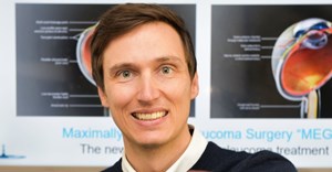
“It is a relatively novel way for surgeons to visualise and assess a patient’s anatomy and can help them to plan and perform procedures with a greater measure of safety and efficiency,” says Dr Rudolph Venter, an orthopaedic surgeon with the FMHS.
Doctors can “recreate” any anatomical structure by importing an image from a CT or MRI image into a software programme, which then enables them to print a plastic 3D model of the image.
3D printing is not a new technology, but has been considered too costly for everyday use until recently. In recent years, however, 3D printers have become much more affordable and the required software is now freely and widely available. This has enabled surgeons to comprehensively explore the potential benefits of using 3D models in medical practice. Orthopaedic surgery is a good place to start this journey of discovery, as the work of the orthopaedic surgeon is very tactile by nature.
According to Venter, orthopaedic procedures require a great deal of pre-operative planning. Historically, orthopaedic surgeons used X-rays and paper templates to plan procedures, but now X-rays, CT scans or MRI images are electronically manipulated to help plan for operations.
“The planning has always been visual, while the actual execution of the procedure is very “hands on”. Having a 3D model of the patient’s anatomy allows you to plan for the operation in a whole new way. For example, not only can you see the tumour you need to remove, but you can also feel it. Or you can physically plan where you are going to make the bone cut to correct a deformity,” explains Venter. He adds that these models can even be used to rehearse interventions or during trial procedures.
“For instance, we would print a patient’s thigh bone with a deformity and rehearse the procedure, or physically experiment with implants in the model as if we were performing the exact procedure on the patient. This enables the surgeon to enter the operating room with confidence, knowing what size implant to use and where to make all the bone cuts,” explains Venter.
3D printed models can also assist surgeons in visualising their patient’s anatomical structures as they are operating. Usually surgery provides only a small “window” into a patient’s body, but the availability of a 3D model of the patient’s own anatomy in theatre can help doctors gauge where to make an incision or position an implant. “For instance, if you have one hand in the wound and one on the model, you obtain a lot of extra information about where the bone fragment or tumour is which you have to remove,” says Venter. “The model becomes a kind of tactile map.”
3D printing enables surgeons to create patient-specific instruments that aid with surgery, assist with training and open up new areas of research. The use of 3D models in medicine is not unique to orthopaedic surgery and can be used in other disciplines as well, including cardiothoracic surgery and neurosurgery.
Venter is currently undertaking research to determine and quantify the advantages of using 3D printed models in day to day orthopaedic practice.